Activity Dependent Plasticity
Current Projects
Cellular activity is the basis of nervous system communication, and it is also a potent tool to alter connections. We target electrical stimulation to the sensorimotor circuits spared by injury in an effort to have them take over the functions of lost connections. An appealing attribute of electrical stimulation is that it can be targeted to adaptive connections to restore function. We have observed short term adaptation of neural circuits to stimulation, which are likely synaptic effects. Electrical stimulation also causes circuits to sprout at their terminations and to form functional connections. These new connections are further reinforced in the setting of repeated practice. Our goal is to better understand the mechanisms of activity-dependent plasticity, so that we can apply in the most effective way for therapy.
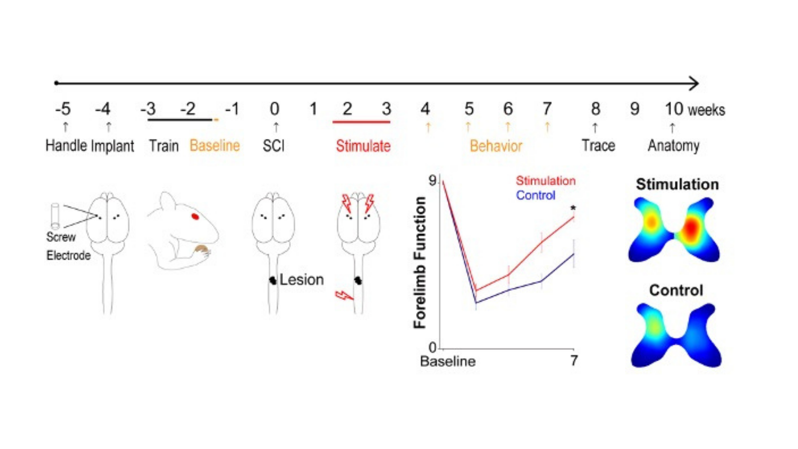
Independent Replication of Motor Cortex and Cervical Spinal Cord Electrical Stimulation to Promote Forelimb Motor Function After Spinal Cord Injury in Rats
Project Leader - QI (Kiki) Yang, PhD
Background: Most patients suffering from the spinal cord injury are injured at cervical level, and majority of them have residual corticospinal tract connections left. We could strengthen these spared connections after incomplete cervical spinal cord injury to improve arm and hand function.
Goal: The goal of this project is to validate independently in our laboratory the rehabilitation effect of paired cortex and spinal cord stimulation protocol developed in Martin Laboratory in rats, as the first step to translate it to human patients use.
Method: We use the combined paradigms of intermittent theta burst stimulation, a mimic version of repetitive TMS, for the cortex and non-invasive transcutaneous direct current stimulation for the spinal cord.
Result: So far, we showed spinal cord injured animals receiving stimulation perform significantly better on behavior tasks challenging their forelimb function than injured animals that received sham treatment. There were also more axons sprouting below the lesion in stimulation rats than controls.
Impact: Our study is a necessary step in establishing a robust therapy, applying a novel treatment to human patients confidently and also strongly endorsed by the spinal cord injury clinical and research community. The large effect size and the replication in independent laboratory validate this approach, which will be trialed in cats before being tested in people using non-invasive methods.
Support: NYS Department of Health Spinal Cord Injury Board C31291GG (JHM)
Paired Motor Cortex and Cervical Spinal Cord Stimulation to Corticospinal Tract Connections and Functional Recovery After Spinal Cord Injury
Project leader - Ajay Pal
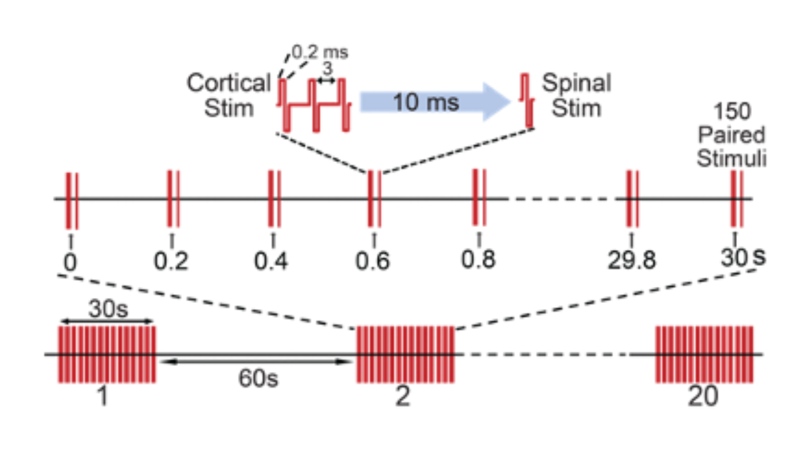
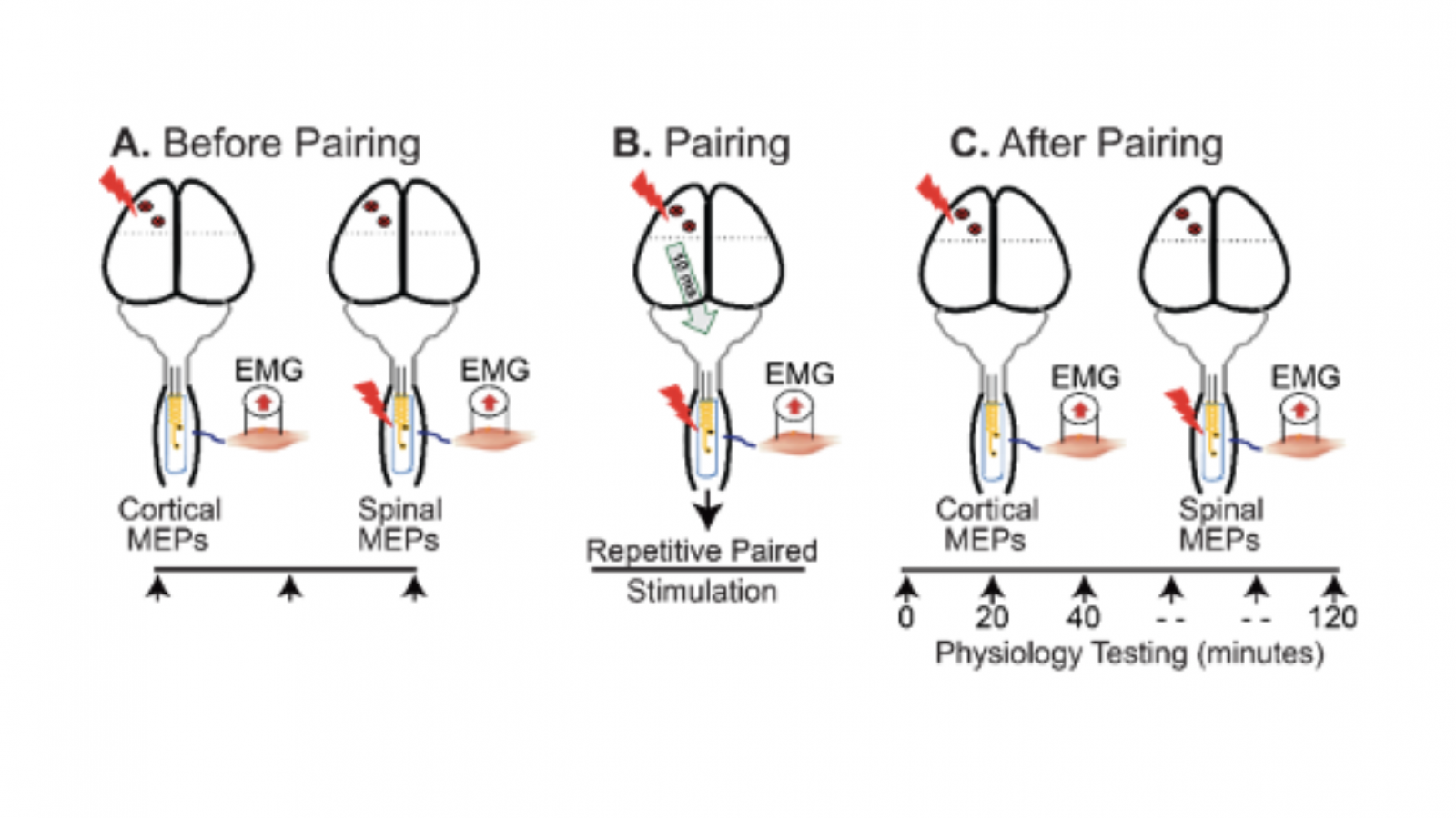
Background: Paralysis from spinal cord injury (SCI) is often the result of damage to the corticospinal tract (CST), the pathway that directly connects motor cortex to the spinal cord. Even in fully paralyzed individuals, however, a portion of the CST is often spared, providing an anatomical substrate for therapeutic interventions. Electrical stimulation of the brain or spinal cord has shown efficacy in improving CST function after SCI, either by amplifying the brain control signals or by increasing the sensitivity of the spinal cord. Whereas paired stimulation of the CST and its spinal targets have the potential to amplify the effects of either alone. The parameters for effective paired stimulation, and the site of their interaction, are not known. Our proposed experiments in rat models of CST damage will define the best location and stimulation parameters for pairing brain and spinal cord stimulation, as a means to improve motor function.
Goal: The goal of our research program is to improve the function of CST circuits and promote recovery of movement after SCI. We will test the hypothesis that pairing motor cortex and spinal cord stimulation will augment CST motor responses and improve forelimb function after SCI.
Methods: To enable safe and effective stimulation, we have developed a novel electrode array, which is inert, thin, and pliable.
Results: In preliminary experiments using this array, we have shown that pairing epidural motor cortex stimulation with epidural cervical spinal cord stimulation results in better potentiation of cortically evoked forelimb muscle responses, than with either on its own. We have also shown that repeating the paired stimulation leads to potentiation of CST responses that persist after stimulation.
Impact: This plasticity occurs at the level of the spinal cord. The experimental evaluation of this innovative paradigm will advance the therapeutic potential of using paired stimulation to enable recovery of function in people with paralysis due to CST damage.
Associated Grant: Paired stimulation in rats
Combining 4-AP with Motor Training to Promote Forelimb Motor Recovery in Rats with Pyramidal Tract Injury
Project Leader - Aditya Ramamurthy, MS
Background: Recovery of forearm movement remains a largely unmet need for cervical spinal cord injury patients. We recently demonstrated that the drug 4-Aminopyridine (4-AP) is capable of raising significant levels of neural excitability from circuits that are spared after injury.
Goal: Our main goal is to optimally combine 4-AP and motor training to produce lasting motor recovery in arm and hand function.
Method: We will assess the changes in supination function associated with paired 4-AP and motor training using a three-pronged approach of behavior testing, anatomic tract tracing and physiological confirmation of which tracts assume control of function after treatment.
Impact: The results from this proposal are poised to provide a new rationale for using an FDA-approved drug which is safe in people with SCI to potentiate the effects of motor training.
Associated Grant: DOH01 PART 2 2017 C33269GG NY State’s Innovative, Developmental or Exploratory Activities in Spinal Cord Injury. Role: Post-Doctoral Fellow, PI Jason Carmel (2018-2020)
Associated Publication: Sindhurakar A, Mishra AM, Gupta D, Iaci JF, Parry TJ, Carmel JB. Clinically relevant levels of 4- Aminopyridine (4-AP) strengthen physiological responses in intact motor circuits in rats, especially after pyramidal tract injury. Neurorehabil Neural Repair. (2017)
Dissecting and Strengthening Corticospinal Connections After Spinal Cord Injury Using Advanced Neuroscience Methods
Project Leader - Ajay Pal, PhD
Background: The motor circuits or tracts that mediate spontaneous recovery in motor function could be strengthened by electrical stimulation to improve functional recover. However, the mechanism underlying spontaneous recovery after spinal cord injury has not been understood.
Goals:
To identify motor tracts and circuits responsible spontaneous recovery after spinal cord injury
To improve functional recovery by strengthening them with electrical stimulation.
Methods:
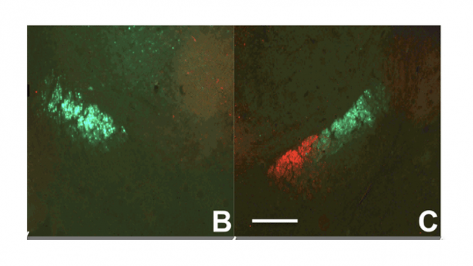
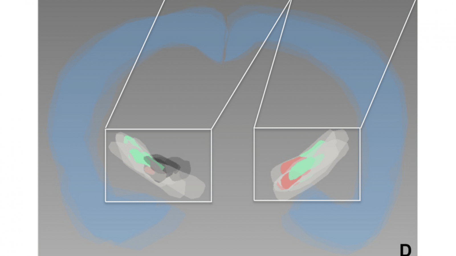
Viral Tracers expressing fluorescent proteins are used for identification of the circuits. Tracer methods and expected results: 1. Anterograde viral tracers are injected into forelimb (FL) motor cortex. Tissue is sectioned coronally at cortex, C4 (lesion site) and C6 spinal cord. Anterogradely labeled axons are represented as dots in the tract and branching lines in the gray matter. The red is not shown in the gray matter. Methods and expected anatomical results: 2. Viruses for inactivation work as anterograde tracer. AAV1-DIO-hM4Di is injected into ipsilateral forelimb motor cortex (FL). AAV9-Cre is injected into C5-C7
Viral inactivation system is used for testing their necessity in spontaneous and electrical stimulation-induced functional recovery after cervical spinal cord contusion injury. Expected behavioral result. Spontaneous functional recovery and enhanced recovery with brain stimulation are degraded by ipsilateral CST inactivation. CNO, circuit specific inactivation with Cre-dependent DREADD and colzapine-N-oxide (CNO) administration; STIM, electrical stimulation.
Results: The efficiency of the viral tracers and viral inactivation system has been tested and confirmed.
Impact: When successfully done, this study will provide information about specific substrates for development of improved therapeutic interventions without off-target effect, that are strengthened to improve functional recovery after spinal cord injury.
Associated grant: Individual Post-Doctoral Award
Contract Number: DOH01-C30859GG-3450000
New York State Department of Health (NYSDOH)
Spinal Cord Injury Research Board (SCIRB)
Associated Publication: Park HG & Carmel J.B. (2016). Selective Manipulation of Neural Circuits. Neurotherapeutics, 13(2), 311-324
Combined Therapy of Forelimb Area Motor Cortex and Spinal Cord Epidural Stimulation to Improve Hand Function After Spinal Cord Injury and Identifying the Responsible Pathway
Project Leader - Ajay Pal, PhD
Background: Residual connections left between the cortex and spinal cord after injury are too weak to result in significant functional recovery. Our lab has shown that paired motor cortex and cervical epidural stimulation convergent in the spinal cord produces long-lasting augmentation in motor evoked potentials that is stronger than using either of them alone. But little is known whether this paired stimulation could further strengthen the weak connections left after injury for hand functional recovery.
Goal: The goal of this study is to use paired forelimb motor cortex and spinal epidural stimulation for the first time to further improve forelimb function after bilateral C4 moderate contusion SCI in rats and identify the pathway responsible for mediating the stimulation effect.
Method: To quantify the proposed therapy’s effect, SCI animals received paired stimulation will be compared vs. animals received sham stimulation on behavior assays of supination knob task and food manipulation (IBB) task. We will inactivate cortico-reticular connection and this is hypothesized to abrogate the effects of paired stimulation.
Predict Result: The hypothesis will be proved true if stimulation animal's performance improves significantly than controls after paired stimulation, and worsens significantly post pathway inactivation compared to itself before inactivation, and to the control animals without inactivation.
Impact: The experiments have the potential to inform our understanding of spared motor connections after cervical spinal cord injury and whether those connections can be recruited using a promising therapeutic approach.
Support: NYS Department of Health Spinal Cord Injury Board C34050GG
Motor Cortex Electrical Stimulation to Augment Spontaneous Recovery After Chronic Subcortical Stroke
Project Leader - Tong-Chen Wen, MD, PhD
Background: Subcortical motor system strokes, such as those that affect the internal capsule and brain stem, are distinct from cortical strokes in that they leave motor cortex and spinal cord intact but destroy axonal connections between them.
Goal: In order to develop effective therapies for this common condition, it is critical to have a reproducible animal model.
Method: We performed electrophysiological motor mapping of the internal capsule and viral tracing fibers emanating from the forelimb area and the hindlimb area of the motor cortex to the internal capsule. We used an optrode, which contains an electrode to localize forelimb responses and an optical fiber to activate a photothrombotic agent with light to ablate the forelimb fibers in the internal capsule.
Results: We found largely separate representations of the forelimb and hindlimb in the internal capsule. This lesion caused lasting impairment in forelimb function, but preserved the hindlimb fibers of the internal capsule.
Impact: The present model induces reproducible white matter damage suitable for studying the pathophysiology of subcortical stroke and for screening the therapeutic strategy of this disease.
Associated grants: NIHR01
Associated publication: Selective lesions to the posterior limb of internal capsule in the rat using microstimulation guidance.