Neural Circuits
Current Projects
The nervous system functions through a network of neural connections. We seek to understand which neural connections are critical for skilled movement, particularly of the arm and hand. We focus on the sensory and motor circuits that connect the brain and spinal cord. Motor connections include the corticospinal tract, the direct connection between cortex and the spinal cord, as well as rely pathways through the brain stem. These motor circuits interact strongly with sensory circuits that code for limb position and muscle length and tension among others. The interplay between descending motor connections and sensory connections is particularly robust in the spinal cord, and we think this interaction may be key for movement in health and for recovery of movement after injury. A key question is which neural circuits should be targeted for recovery. To address this, we have used viral methods to label and manipulate specific neural connections to understand their role in endogenous recovery after injury. We also use targeted electrical stimulation to promote specific connections, and particularly the interaction of sensory and motor connections in the spinal cord. The overall approach is to develop an understanding of the neural networks that should be recruited for nervous system repair.

Neural Circuits
Dissecting and Strengthening Corticospinal Connections After Spinal Cord Injury Using Advanced Neuroscience Methods
Project Leader - Ajay Pal, PhD
Background: The motor circuits or tracts that mediate spontaneous recovery in motor function could be strengthened by electrical stimulation to improve functional recover. However, the mechanism underlying spontaneous recovery after spinal cord injury has not been understood.
Goals
- To identify motor tracts and circuits responsible spontaneous recovery after spinal cord injury
- To improve functional recovery by strengthening them with electrical stimulation.
Methods
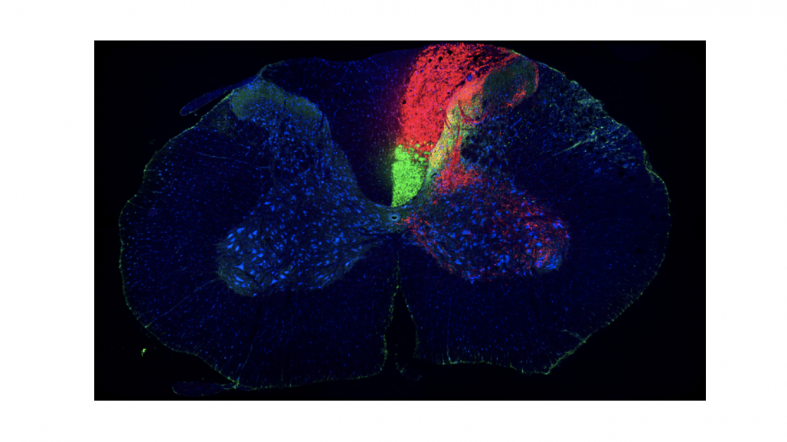
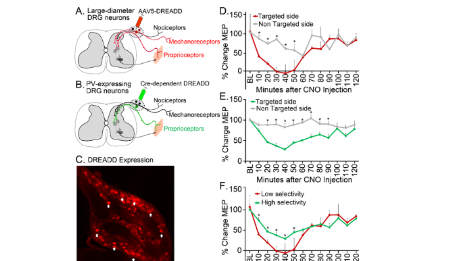
- Viral Tracers expressing fluorescent proteins are used for identification of the circuits. Tracer methods and expected results: 1. Anterograde viral tracers are injected into forelimb (FL) motor cortex. Tissue is sectioned coronally at cortex, C4 (lesion site) and C6 spinal cord. Anterogradely labeled axons are represented as dots in the tract and branching lines in the gray matter. The red is not shown in the gray matter. Methods and expected anatomical results 2. Viruses for inactivation work as anterograde tracer. AAV1-DIO-hM4Di is injected into ipsilateral forelimb motor cortex (FL). AAV9-Cre is injected into C5-C7
- Viral inactivation system is used for testing their necessity in spontaneous and electrical stimulation-induced functional recovery after cervical spinal cord contusion injury. Expected behavioral result. Spontaneous functional recovery and enhanced recovery with brain stimulation are degraded by ipsilateral CST inactivation. CNO, circuit specific inactivation with Cre-dependent DREADD and colzapine-N-oxide (CNO) administration; STIM, electrical stimulation.
Results: The efficiency of the viral tracers and viral inactivation system has been tested and confirmed.
Impact: When successfully done, this study will provide information about specific substrates for development of improved therapeutic interventions without off-target effect, that are strengthened to improve functional recovery after spinal cord injury.
Associated grant: Individual Post-Doctoral Award
Contract Number: DOH01-C30859GG-3450000
New York State Department of Health (NYSDOH)
Spinal Cord Injury Research Board (SCIRB)
Associated Publication: Park HG & Carmel J.B. (2016). Selective Manipulation of Neural Circuits. Neurotherapeutics, 13(2), 311-324
Repairing Sensorimotor Connections After Neonatal Lesion
Project Leader - Tong-Chen Wen MD, PhD
Background: The young brain is more plastic in its connections than older animals. As a result, animals with nervous system injury early in life often recovery significant function.
Goal: The overall goal of our experiments is to understand how in neonatal animals brain plasticity allows connections between the brain and spinal cord to become strong after injury. We will also try to improve on this natural recovery by electrically stimulating the brain-spinal cord connections that are spared by injury.
Methods: We used neural tracers to test plasticity and behavioral tasks to investigate motor deficits after neonatal pyramidotomy.
Results: We found that the uninjured hemisphere may encode separate control of the unimpaired and the impaired forelimbs of rats with neonatal injury.
Impact: This will help us to understand why animals recover better after early injury versus later injury. It will also test whether we can improve on the natural recovery process. This research may have direct relevance for humans with early brain injury.
Associated grants: This study was supported by funding from National Institutes of Health (NIH)-National Institute of Neurological Disorders and Stroke (NINDS) K08 NS073796 and The Thomas and Agnes Carvel Foundation.
Associated publication: Plasticity in One Hemisphere, Control From Two: Adaptation in Descending Motor Pathways After Unilateral Corticospinal Injury in Neonatal Rats.